7 Times You Should Get a Mole Checked Even If It Looks Normal

Heading 2

Lorem ipsum dolor sit amet, consectetur adipisicing elit. Nostrum minus ea suscipit porro alias corporis libero at. Perferendis omnis, veniam nemo beatae vel? Tempora numquam a repellat eaque natus, magnam?

Heading 2

Lorem ipsum dolor sit amet, consectetur adipisicing elit. Autem ipsum mollitia neque, illum illo excepturi, eum incidunt fugit nostrum est, voluptate eaque minima corporis debitis at, dolores ipsam. Quaerat, dolores.

Heading 2

Lorem ipsum dolor sit amet, consectetur adipisicing elit. Autem ipsum mollitia neque, illum illo excepturi, eum incidunt fugit nostrum est, voluptate eaque minima corporis debitis at, dolores ipsam. Quaerat, dolores.

Heading 2

Lorem ipsum dolor sit amet, consectetur adipisicing elit. Autem ipsum mollitia neque, illum illo excepturi, eum incidunt fugit nostrum est, voluptate eaque minima corporis debitis at, dolores ipsam. Quaerat, dolores.

Heading 2

Lorem ipsum dolor sit amet, consectetur adipisicing elit. Autem ipsum mollitia neque, illum illo excepturi, eum incidunt fugit nostrum est, voluptate eaque minima corporis debitis at, dolores ipsam. Quaerat, dolores.

Most people rely on the ABCDE rule (Asymmetry, Border, Color, Diameter, Evolution) when scanning their skin for trouble. It’s a helpful starting point, but it doesn’t catch everything, and some concerning moles never break a single one of those rules. So, relying on appearance alone can leave real risks unnoticed. 

Keep reading to learn about seven situations where a professional mole check is worth scheduling, even if the mole in question looks completely unremarkable.
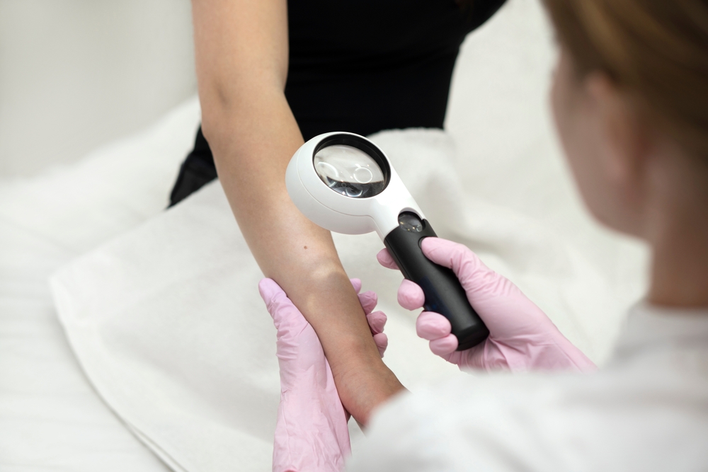
Examining a mole

Can a Mole Look Normal and Still Be a Problem?

It’s a common misconception that dangerous moles always look obviously abnormal. Some forms of melanoma do not present with the classic warning signs most people are taught to watch for.

For example, amelanotic melanomas can appear pink, red, or skin colored rather than dark and irregular, and nodular melanomas tend to grow as a firm, evenly colored bump that can easily be mistaken for a harmless cyst or pimple.

This is part of why dermatologists don’t just rely on visual pattern recognition. 

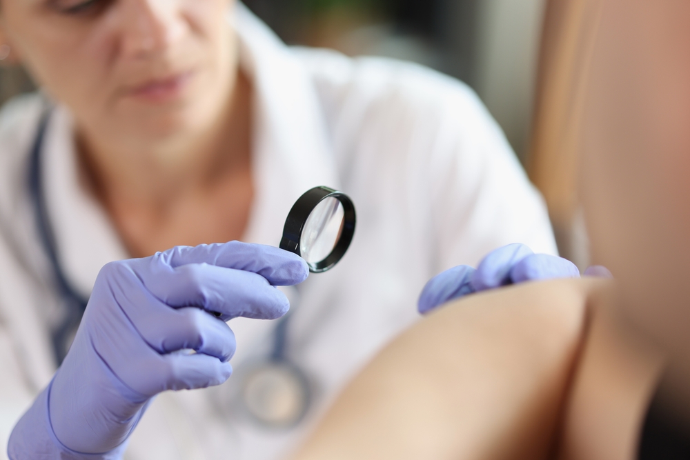
A trained provider at Calkin & Boudreaux uses dermoscopy and clinical experience to evaluate moles that might otherwise be overlooked by the untrained eye.

1. When You Have a New Mole After Age 30

Most moles form during childhood and adolescence, with the total count typically leveling off by early adulthood. A new mole that appears after age 30 is statistically less common and deserves attention, even if it looks small and unremarkable. While this doesn’t mean every new mole after 30 is dangerous, the odds shift enough that a quick evaluation is a reasonable precaution rather than an overreaction.

2. When a Mole Has Changed, Even Subtly

Dramatic changes tend to grab attention right away, but subtle shifts are just as important to track. A slight change in texture, a faint new shade within the mole, or a small increase in size over several months can all be early signals worth investigating.

Photographing your moles every few months can help you notice these gradual differences that might otherwise slip by unnoticed.

Joel Crockett, MD a board-certified dermatologist at Golden State Dermatology in Walnut Creek and San Ramon, explains why subtle change matters so much:

“Patients often assume that only dramatic changes are worth mentioning, but some of the most important findings I make are small ones. A mole that has gone from perfectly flat to just slightly raised, or one that has developed a faint new color within it, can be an early clue that something is evolving. Catching these changes early is exactly why we recommend regular skin checks rather than waiting for something to look obviously wrong.”

Trusting your own sense that something is different, even without a clear reason why, is valid. It’s often worth a conversation with your provider.

3. When You Have a Personal History of Skin Cancer

Patients who have previously been treated for melanoma or other skin cancers face a higher chance of developing new lesions elsewhere on the body.  Regular surveillance becomes especially important in these cases, since early detection dramatically improves outcomes. Your provider at Calkin & Boudreaux can set up a personalized follow-up schedule based on your specific history and risk factors.


4. When You Have a Family History of Melanoma or Skin Cancer

Genetics play a large role in skin cancer risk. If a parent, sibling, or child has been diagnosed with melanoma, your own risk increases, which lowers the threshold for what’s worth having checked.

Family history can also mean your provider recommends more frequent skin exams than someone without that background. Sharing your full family history during appointments helps your dermatologist tailor a monitoring schedule that fits your actual risk level.

5. When the Mole Is in a Hard-to-See Spot

The back, scalp, backs of the ears, and areas between the toes are all commonly missed during a typical mirror check. A professional full-body skin exam looks at these hard-to-see spots where a problematic mole could be easy to miss. A dermatologist can examine these areas that are difficult or impossible to see on your own, using proper lighting and tools designed for thorough evaluation.

Michelle Cheng, MD, a board-certified dermatologist at Golden State Dermatology in Torrance, notes how often this comes up in practice: 

“I can’t tell you how many times I’ve found something significant in a spot the patient had never even considered checking, like the scalp or the sole of the foot. These areas don’t get regular sun exposure in some cases, but they’re still at risk, and patients simply can’t monitor them well on their own. It’s one of the strongest reasons to come in for a full body exam rather than relying only on what you can see in a mirror.”

Asking a partner, family member, or friend to help check these areas at home can also be useful between professional visits.

6. When You’ve Had Significant Sun Exposure or Tanning Bed Use

Cumulative UV exposure over a lifetime raises baseline skin cancer risk, regardless of how any individual mole currently looks. This applies to both prolonged sun exposure and indoor tanning bed use, even if that exposure happened decades ago. If this sounds like your background, it’s worth mentioning to your provider so they can factor it into your recommended screening schedule.
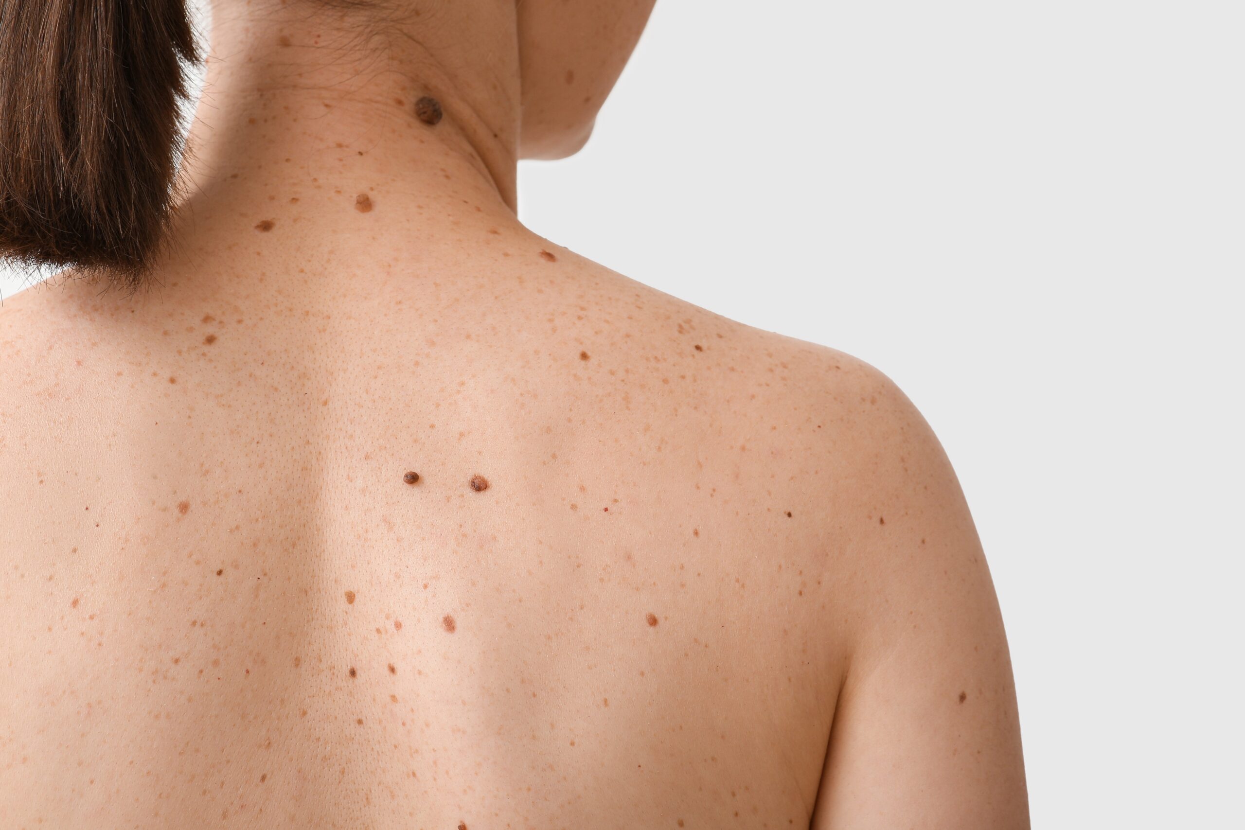
A large mole

7. When You Notice Symptoms Like Itching, Bleeding, or Tenderness

Visual changes aren’t the only signal worth paying attention to. A mole that itches persistently, bleeds without an obvious cause, or feels tender to the touch can be flagging a problem before any visible change appears.

These symptoms are easy to dismiss, especially if the mole still looks the same as it always has. Reporting them to a dermatologist ensures they get properly evaluated rather than ignored.

Why Annual Skin Checks Matter Regardless of Appearance

The situations above all share the common thread that none of them depend on a mole looking obviously dangerous. Risk factors like age, family history, sun exposure, and mole location matter just as much as visual appearance, sometimes more.

This is exactly why annual skin exams are recommended even for patients who feel confident in their own self-checks. A professional evaluation combines clinical training, proper tools, and a full body view that’s simply impossible to replicate at home.

Calkin & Boudreaux providers are trained to catch the findings that don’t fit the typical warning signs, giving patients an added layer of protection beyond what the ABCDE rule alone can offer. Consistent, proactive screening remains one of the most effective tools for catching skin cancer early.

If you have questions about your skin, schedule an appointment at Calkin & Boudreaux today!
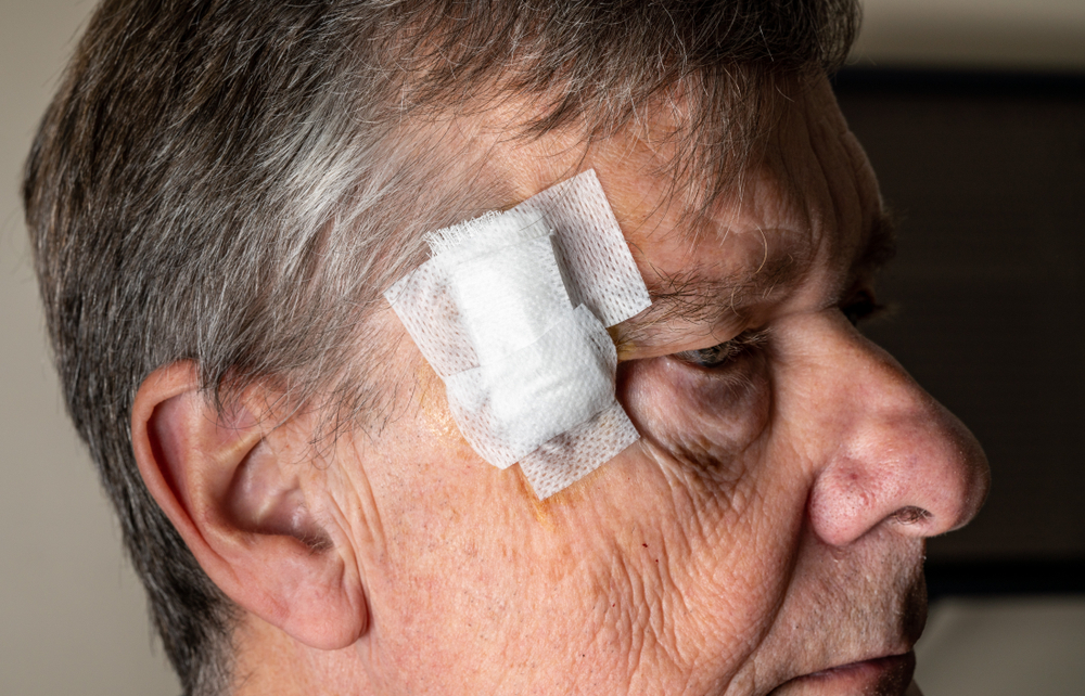
Can You Speed Up Healing After Mohs?

Heading 2

Lorem ipsum dolor sit amet, consectetur adipisicing elit. Nostrum minus ea suscipit porro alias corporis libero at. Perferendis omnis, veniam nemo beatae vel? Tempora numquam a repellat eaque natus, magnam?

Heading 2

Lorem ipsum dolor sit amet, consectetur adipisicing elit. Autem ipsum mollitia neque, illum illo excepturi, eum incidunt fugit nostrum est, voluptate eaque minima corporis debitis at, dolores ipsam. Quaerat, dolores.

Heading 2

Lorem ipsum dolor sit amet, consectetur adipisicing elit. Autem ipsum mollitia neque, illum illo excepturi, eum incidunt fugit nostrum est, voluptate eaque minima corporis debitis at, dolores ipsam. Quaerat, dolores.

Heading 2

Lorem ipsum dolor sit amet, consectetur adipisicing elit. Autem ipsum mollitia neque, illum illo excepturi, eum incidunt fugit nostrum est, voluptate eaque minima corporis debitis at, dolores ipsam. Quaerat, dolores.

Heading 2

Lorem ipsum dolor sit amet, consectetur adipisicing elit. Autem ipsum mollitia neque, illum illo excepturi, eum incidunt fugit nostrum est, voluptate eaque minima corporis debitis at, dolores ipsam. Quaerat, dolores.

Mohs surgery is one of the most effective treatments available for many types of skin cancer, offering high cure rates while preserving as much healthy tissue as possible. Once the procedure is complete, most patients want to know how they can heal as quickly and smoothly as possible.

While no single trick can shrink your body’s natural healing timeline overnight, there are steps you can take to support recovery and reduce the risk of complications. Keep reading to learn what actually helps your skin heal after Mohs surgery, and what to watch for along the way.

What Is Mohs Surgery?

Mohs surgery is a precise surgical technique used to remove skin cancer layer by layer. Each layer is examined under a microscope in real time, allowing your surgeon to confirm that all cancerous cells have been removed before closing the wound.


This method offers one of the highest cure rates of any skin cancer treatment, mainly because it allows for a thorough examination of the tissue margins.

Because Mohs surgery can involve removing more than one layer of tissue, the size of the resulting wound can vary quite a bit from patient to patient.

Your provider at Calkin & Boudreaux will discuss your specific wound and repair method before you leave your appointment.

What Does Normal Healing After Mohs Look Like?

Every patient’s healing process looks a little different depending on the size and location of the wound. That said, most people can expect some redness, mild swelling, and scabbing in the days following surgery.

In the first week, it’s common for the area to feel tender or slightly itchy as new tissue begins to form. Swelling and bruising often peak within the first two to three days before gradually improving.

Over the next several weeks, the wound will continue to close and the surrounding skin will slowly return to a more normal appearance. Complete healing, including the fading of redness and refinement of any scar, can take several months to a full year depending on the treatment area.

How Can You Care for the Wound Site?

Proper wound care is one of the most important factors in how well and how quickly your skin heals.

Keeping the area clean and appropriately moist, rather than letting it dry out and scab excessively, tends to support faster healing with less visible scarring.

Your provider will typically recommend gently cleaning the wound with mild soap and water, followed by an application of a bland ointment such as petroleum jelly. Covering the area with a bandage as instructed also helps protect it from bacteria and reduces the risk of irritation from clothing or accidental bumps.

Jeanne Strunck, MD, a board-certified dermatologist and Mohs surgeon at Golden State Dermatology, explains why consistent wound care matters so much.

“Patients are often surprised to learn that keeping a wound slightly moist actually helps it heal faster and with a better cosmetic outcome than letting it air out and scab over. Scabs can actually slow down the migration of new skin cells across the wound bed. We give very specific instructions for a reason, and following them closely in those first one to two weeks can make a noticeable difference in the final result.”

Does Nutrition Play a Role in Healing?

What you eat and drink in the weeks following surgery can influence how your body repairs itself.

Staying well hydrated supports healthy circulation, which helps deliver oxygen and nutrients to the healing tissue. Protein is particularly important during this time as well, since it provides the building blocks your body uses to repair skin. Foods rich in vitamin C and zinc, such as citrus fruits, leafy greens, and lean meats, can also support the healing process.

On the flip side, alcohol and smoking can both interfere with healing by affecting circulation and collagen production. Avoiding or significantly limiting these during your recovery period is one of the simplest ways to give your skin a better chance at healing efficiently.

Can Activity Level Affect Recovery?

A doctor examining a patient

It’s tempting to jump right back into your normal routine, but overdoing it too soon can slow healing or even affect your scar’s appearance. Strenuous exercise, heavy lifting, and activities that stretch or strain the surgical site should generally be avoided for at least the first one to two weeks. Gentle movement is usually fine and even encouraged to support circulation. Your provider will give you specific guidance based on the location of your wound and the type of repair that was performed.

Are There Products That Help (or Hurt) Healing?

Once your wound has closed, certain products can help minimize the appearance of your scar over time.

For example, silicone-based gels and sheets are among the most well-researched options for improving scar texture and color.

Broad-spectrum sunscreen is also essential once the area is healed enough to apply it, since new scar tissue is especially vulnerable to sun damage and discoloration.

Your provider at Calkin & Boudreaux can recommend a timeline for introducing these products based on your specific healing progress.

Katie Given, MD, PhD, a board-certified dermatologist and Mohs surgeon at Golden State Dermatology, cautions against reaching for unproven remedies.

“Patients often ask about home remedies they’ve seen online, like vitamin E oil or various essential oils, but the evidence behind many of these is thin at best. Some can even irritate healing skin or trigger an allergic reaction. I always recommend sticking to what your provider has specifically approved rather than experimenting on your own during this sensitive healing window.”

When Should You Contact Your Provider?

While some redness, swelling, and mild discomfort are expected, certain symptoms warrant a call to your provider. Increasing pain, spreading redness, or warmth around the wound can all be signs of infection.

A fever, unusual discharge, or a wound that seems to be opening rather than closing are also reasons to reach out promptly.

Trusting your instincts and checking in with your provider is always better than waiting to see if something resolves on its own.

Supporting Your Skin’s Natural Healing Process

Healing after Mohs surgery is a gradual process that can’t be rushed dramatically, but the choices you make in the days and weeks afterward do matter. Careful wound care, good nutrition, appropriate activity levels, and sun protection all work together to support your skin as it repairs itself. Following your personalized aftercare instructions closely gives your skin the best possible chance at a smooth and successful healing process.

If you have questions about your skin, schedule an appointment at Calkin & Boudreaux today!
What Is the Difference Between a Dermatopathologist and a Regular Pathologist

Heading 2

Lorem ipsum dolor sit amet, consectetur adipisicing elit. Nostrum minus ea suscipit porro alias corporis libero at. Perferendis omnis, veniam nemo beatae vel? Tempora numquam a repellat eaque natus, magnam?

Heading 2

Lorem ipsum dolor sit amet, consectetur adipisicing elit. Autem ipsum mollitia neque, illum illo excepturi, eum incidunt fugit nostrum est, voluptate eaque minima corporis debitis at, dolores ipsam. Quaerat, dolores.

Heading 2

Lorem ipsum dolor sit amet, consectetur adipisicing elit. Autem ipsum mollitia neque, illum illo excepturi, eum incidunt fugit nostrum est, voluptate eaque minima corporis debitis at, dolores ipsam. Quaerat, dolores.

Heading 2

Lorem ipsum dolor sit amet, consectetur adipisicing elit. Autem ipsum mollitia neque, illum illo excepturi, eum incidunt fugit nostrum est, voluptate eaque minima corporis debitis at, dolores ipsam. Quaerat, dolores.

Heading 2

Lorem ipsum dolor sit amet, consectetur adipisicing elit. Autem ipsum mollitia neque, illum illo excepturi, eum incidunt fugit nostrum est, voluptate eaque minima corporis debitis at, dolores ipsam. Quaerat, dolores.

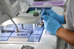
A skin biopsy may feel like the main event, but the real diagnostic work is just beginning. Once your sample leaves the procedure room, it heads to a lab where someone examines it under a microscope to determine exactly what is going on. That someone is a pathologist, but not all pathologists are the same when it comes to skin.

Keep reading to learn the difference between a general pathologist and a dermatopathologist, and why it matters for your diagnosis.

What Is a Pathologist?

A pathologist is a physician who specializes in diagnosing disease by examining tissue, blood, and other body fluids. Their work happens largely behind the scenes, but their reports directly shape the treatment decisions your other doctors make.

General pathologists are trained broadly across the entire body. This wide-ranging expertise is essential to modern medicine. However, the breadth of their work means they don’t always have the deep, narrow focus that certain organ systems demand.

What Is a Dermatopathologist?

A dermatopathologist is a physician who has completed additional, subspecialty training focused exclusively on diseases of the skin, hair, and nails. They look at skin samples all day, every day, which gives them a particularly refined eye for what is and isn’t normal.

Dermatopathologists are board-certified in dermatopathology, a credential earned only after years of additional fellowship training. Their training equips them to recognize hundreds of skin conditions, including rare disorders that a general pathologist may rarely encounter.

Dr. Michelle Cheng, a board-certified dermatologist at Golden State Dermatology in Torrance, explains:

“The difference between a general pathologist and a dermatopathologist often comes down to volume and focus. A dermatopathologist may read tens of thousands of skin biopsies over the course of their training and career, and that kind of repetition sharpens diagnostic accuracy in ways that are hard to overstate. When we send a biopsy out, we want it interpreted by someone who has seen the subtle variations of that exact condition many times before. For our patients, that focused expertise can make a real difference in catching things early and avoiding misdiagnosis.”
Why Skin Is Uniquely Complex Under the Microscope

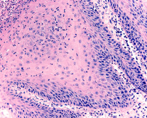
Skin may seem like a simple organ from the outside, but under a microscope, it is remarkably intricate. It has multiple layers, each with its own cell types, structures, and patterns that can be affected by disease in different ways.

Many skin conditions also look surprisingly similar at first glance. A benign mole and an early melanoma can share features, certain rashes can mimic skin cancers, and inflammatory conditions often overlap in their microscopic appearance. Recognizing a melanoma in its earliest stages, for example, can depend on noticing subtle architectural changes that a less experienced eye might overlook. The same is true for distinguishing one type of lymphoma from another or identifying an unusual presentation of an autoimmune condition.

Dermatopathologists are trained specifically to spot these subtleties. Their pattern recognition is built on years of focused exposure to skin tissue rather than tissue from across the body.

When Does a Skin Biopsy Get Read by a Dermatopathologist?

Not every skin biopsy in the country is automatically reviewed by a dermatopathologist. Where your sample is sent often depends on the dermatology practice, the laboratory they partner with, and sometimes your insurance plan.

Dermatology practices that prioritize accurate skin diagnoses often work directly with dermatopathology labs or in-house dermatopathologists. This intentional choice helps ensure that the physician interpreting your tissue has the specific expertise that skin samples require.

If you are ever curious about who reads your biopsies, it is completely reasonable to ask your dermatologist. Knowing the answer can give you greater confidence in your results, especially when the diagnosis is something serious.

How Accuracy Affects Your Diagnosis and Treatment

Dermatopathologists tend to have higher diagnostic accuracy for skin samples than general pathologists, particularly for melanocytic lesions such as moles and melanomas. That accuracy translates directly into better treatment decisions and better outcomes.

Sarah Tadros, PA-C, a certified physician assistant at Golden State Dermatology in Montclair and Glendora, adds:

“In dermatology, the line between a benign growth and something dangerous can be incredibly fine on a slide. We rely heavily on dermatopathologists to draw that line correctly, because the next step in a patient’s care depends entirely on what the biopsy report says. A clear, accurate diagnosis allows us to recommend the right treatment with confidence, whether that means continued monitoring, a minor procedure, or a more involved surgical plan. The value of that expertise is hard to overstate when you are talking about skin cancer in particular.”

When a dermatopathologist reads your biopsy, your dermatologist receives a report shaped by specialized expertise. That report often includes specific details about the depth, type, and behavior of any abnormal cells.

These details help your dermatologist tailor your treatment plan with precision. They also help guide follow-up recommendations, such as how often you should be checked for new or changing lesions in the future.

What This Means for Patients at Calkin & Boudreaux Dermatology Associates

Calkin & Boudreaux Dermatology Associates works with Golden State Dermatopathology, a full-service dermatopathology lab based in Walnut Creek, California, designed to deliver fast turnaround times, efficient communication, and advanced diagnostics for both patients and physicians.

That relationship has real benefits for patients. Every case submitted to Golden State Dermatopathology is reviewed by an experienced dermatopathologist, and the lab is led by a team of board-certified specialists with decades of combined experience.

Direct Collaboration Between Your Dermatologist and the Lab

Because both groups work hand in hand, your dermatologist can consult directly with the dermatopathologist reading your biopsy. That kind of open communication is uncommon in outside lab arrangements, and it leads to more accurate diagnoses and more tailored treatment plans.

Fast Turnaround for Faster Answers

Waiting for biopsy results is one of the most stressful parts of any skin procedure. Golden State Dermatopathology offers a typical 1 to 2 day turnaround, which means faster answers and, when needed, faster treatment for serious conditions like melanoma.

Advanced Diagnostic Tools When Cases Get Complex

Not every biopsy is straightforward. For challenging cases, the lab offers advanced services like immunohistochemistry, molecular testing, and FISH testing to distinguish between conditions that can look nearly identical under a routine microscope.

Choosing a dermatology practice that values dermatopathology expertise is one of the best things you can do for your skin health. The right eyes on your slide can mean the difference between an early catch and a missed diagnosis, between confident treatment and unnecessary uncertainty.

If you have questions about your skin, schedule an appointment at Calkin & Boudreaux Dermatology Associates today!

Is Melanoma Rough to the Touch?

Heading 2

Lorem ipsum dolor sit amet, consectetur adipisicing elit. Nostrum minus ea suscipit porro alias corporis libero at. Perferendis omnis, veniam nemo beatae vel? Tempora numquam a repellat eaque natus, magnam?

Heading 2

Lorem ipsum dolor sit amet, consectetur adipisicing elit. Autem ipsum mollitia neque, illum illo excepturi, eum incidunt fugit nostrum est, voluptate eaque minima corporis debitis at, dolores ipsam. Quaerat, dolores.

Heading 2

Lorem ipsum dolor sit amet, consectetur adipisicing elit. Autem ipsum mollitia neque, illum illo excepturi, eum incidunt fugit nostrum est, voluptate eaque minima corporis debitis at, dolores ipsam. Quaerat, dolores.

Heading 2

Lorem ipsum dolor sit amet, consectetur adipisicing elit. Autem ipsum mollitia neque, illum illo excepturi, eum incidunt fugit nostrum est, voluptate eaque minima corporis debitis at, dolores ipsam. Quaerat, dolores.

Heading 2

Lorem ipsum dolor sit amet, consectetur adipisicing elit. Autem ipsum mollitia neque, illum illo excepturi, eum incidunt fugit nostrum est, voluptate eaque minima corporis debitis at, dolores ipsam. Quaerat, dolores.

When people notice a new or changing spot on their skin, one of the first things they do is touch it. If something feels different, raised, or rough, it’s a natural warning sign. But relying on texture to identify melanoma is not as reliable as you might think.

Melanoma, the most serious form of skin cancer, can feel smooth, rough, raised, or completely flat, and sometimes there’s no noticeable difference at all compared to surrounding skin.

Keep reading to learn what melanoma actually feels and looks like, what warning signs to watch for, and why a dermatologist’s eye is always worth more than a fingertip check!

Continue reading “Is Melanoma Rough to the Touch?” →